

For decades, one of the biggest challenges in biology has been simple to describe but incredibly difficult to solve: How do you observe living cells in extreme detail without disturbing them?
Scientists have long relied on fluorescent dyes and chemical labels to make tiny cellular structures visible under microscopes. These methods have led to major discoveries, but they come with a problem. The labels themselves can interfere with natural biological processes.
Some fluorescent molecules fade over time. Others may alter the behavior of the structures they are supposed to observe. And prolonged imaging can damage living cells. Now, researchers at Stanford University have developed a revolutionary microscope that may solve many of these problems at once.
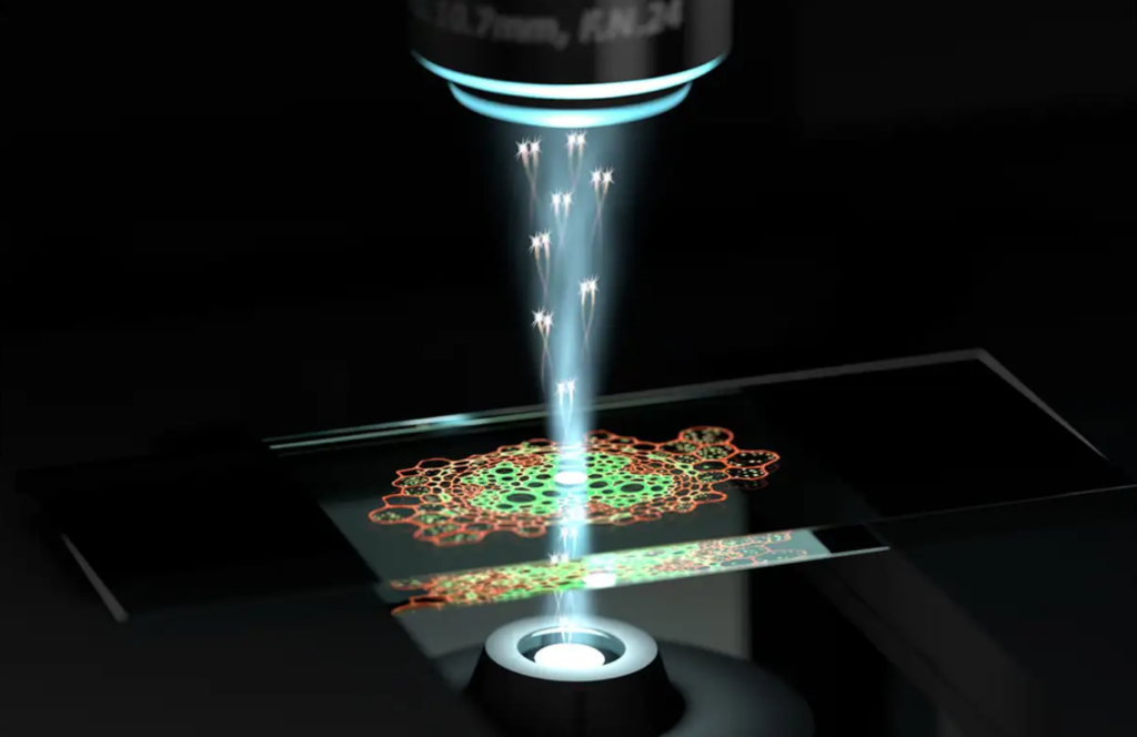
The new system, called Interferometric Image Scanning Microscopy, can observe living cells at an extraordinary resolution of around 120 nanometers without requiring fluorescent labels. Scientists say it is the highest-resolution label-free imaging technique ever demonstrated for live-cell microscopy. Many researchers believe this breakthrough could transform how scientists study diseases, drug interactions, infections, and the fundamental machinery of life itself.
Why Is Looking Inside Cells So Difficult?
Cells are incredibly small. Most of the important structures inside them are measured in nanometers.
For comparison:
- A human hair is about 80,000–100,000 nanometers wide.
- Many cellular structures are only a few hundred nanometers across.
- Some molecular systems are even smaller.
Traditional optical microscopes face a major limitation called the diffraction limit. Because light behaves like a wave, extremely tiny objects begin to blur together. For over a century, this limited how much detail scientists could observe using ordinary light microscopy.
Researchers developed various super-resolution techniques to overcome this barrier, but many of them depend heavily on fluorescent labeling and complicated sample preparation.
What Makes Stanford’s Microscope Different?
The Stanford team wanted to create a microscope capable of observing living cells in their natural state. Instead of attaching glowing labels to cellular components, they developed a system that can directly detect tiny structures using the way light scatters from them.
This means researchers can watch biological activity unfold naturally without constantly modifying or tagging the cell. The result is a much more realistic picture of what is actually happening inside living systems.
Scientists can now observe:
- moving organelles,
- intracellular transport,
- membrane dynamics,
- structural changes,
- and cellular responses
for much longer periods than before.
The Science Behind iISM :
The breakthrough comes from combining two advanced microscopy methods into a completely new imaging system. The first technique is called Interferometric Scattering Microscopy (iSCAT). This method detects extremely tiny objects by measuring how they scatter light.
Rather than relying on fluorescent glow, it analyzes subtle changes in light waves interacting with cellular structures. The second technique is called Image Scanning Microscopy (ISM).
ISM improves resolution by collecting multiple perspectives of the same location and reconstructing a sharper image. Stanford researchers combined these approaches into a hybrid system known as Interferometric Image Scanning Microscopy (iISM). (www.nature.com) This combination dramatically increases image clarity while maintaining the ability to observe living cells without labels.
How Does It Actually Work?
Imagine trying to observe a city from space. One camera provides a single image. But if hundreds of cameras capture slightly different viewpoints simultaneously, powerful software can combine all of those perspectives into a much clearer picture.
The iISM microscope works similarly :-
Instead of using a single detector, it uses an advanced detector array that captures many views of the same microscopic region. Researchers then use specialized algorithms to combine those measurements into highly detailed images.
Stanford scientists describe it as moving from seeing with two eyes to seeing with hundreds of eyes at once. The result is a much sharper view of structures inside living cells.
Why Is 120 Nanometers Such a Big Deal?
Resolution determines how close two objects can be before they appear blurred together. The smaller the number, the finer the detail scientists can observe. Stanford’s system achieved approximately 120-nanometer lateral resolution while remaining label-free.
That level of detail allows researchers to observe:
- endoplasmic reticulum tubules,
- intracellular vesicles,
- cytoskeletal structures,
- membrane behavior,
- and many other microscopic cellular components.
Importantly, researchers can watch these structures moving in real time. Instead of taking static snapshots, scientists can observe dynamic biological processes as they happen.
Why Is Label-Free Imaging So Important?
Fluorescent microscopy has been one of biology’s most powerful tools. However, it comes with limitations. Scientists often need to genetically modify cells or introduce fluorescent chemicals.
These labels can:
- wear out over time,
- alter biological behavior,
- create experimental artifacts,
- and limit observation duration.
The Stanford microscope avoids many of these problems. Because no fluorescent labels are required, cells remain closer to their natural state. Researchers can study biological systems with fewer disturbances.
This is especially important when investigating sensitive processes such as:
- drug interactions,
- infections,
- immune responses,
- and cellular stress mechanisms.
Another Major Advantage:
Less Cell Damage Light itself can damage living cells. This problem is known as phototoxicity. Many high-resolution imaging methods require intense illumination that can stress or harm biological samples.
The Stanford team designed iISM to operate at substantially lower illumination power than many comparable imaging approaches. This means researchers can watch cells for longer periods without significantly disturbing them. Long-term observation is critical because many biological processes occur over extended timescales.
What Could This Mean for Medicine?
The medical implications are enormous. Scientists are already exploring how iISM can be used to study:
- cancer drug uptake,
- pathogen interactions,
- malaria infections,
- cellular transport systems,
- and host-microbe relationships.
Researchers can now observe how cells react to treatments in unprecedented detail. This may help scientists understand why certain drugs succeed while others fail.
Future applications could improve:
- cancer research,
- infectious disease studies,
- personalized medicine,
- and drug development.
Could This Change Biological Research?
Many scientists believe it could. One reason is that biology often depends on observing systems exactly as they behave naturally. The less researchers interfere with a cell, the more accurate their observations become.
The iISM microscope provides something researchers have wanted for years:
high-resolution imaging with minimal disruption. This opens new possibilities for studying living systems in near-native conditions.
What Happens Next?
The Stanford team is already working on improving the technology and expanding its availability.
Researchers hope future versions will provide:
- even higher resolution,
- faster imaging speeds,
- larger fields of view,
- and broader compatibility with existing microscopy systems.
Scientists also plan to combine iISM with fluorescence microscopy. This hybrid approach could provide both molecular specificity and label-free contextual imaging simultaneously.
The Bigger Picture :
For centuries, biology has depended on improving our ability to see. Every major leap in microscopy has revealed entirely new worlds hidden inside living organisms. The invention of the microscope exposed cells.
Electron microscopes revealed molecular structures. Super-resolution imaging broke long-standing physical limits. Now Stanford’s iISM technology offers something different: the ability to watch living cells operate in extraordinary detail while remaining largely undisturbed.
Instead of forcing biology to glow under artificial labels, scientists can simply watch life unfold as it naturally happens. And that may lead to some of the most important discoveries yet.
Sources :
Stanford Research News – iISM Microscope Breakthrough
Light: Science & Applications Research Paper
Phys.org Coverage of the Stanford Microscope